THE BONNER METHOD
Breakthrough Treatment for Periodontitis
Over half of the world's population suffers from periodontal disease. These diseases mainly cause bad breath, bleeding gums and tooth loosening. However, a study conducted in 2014 by dental surgeon Dr. Mark Bonner confirms the systematic presence of pathogenic oral parasites in periodontal diseases; the elimination of these parasites would therefore promote a permanent cure of periodontal diseases.
Dr. Bonner's method for curing periodontal disease, known as the "Bonner Method", consists of microscopically controlled antiparasitic treatment. This method consists in particular in setting up working tools for the diagnosis of periodontal diseases and the restoration of a healthy microbial flora. Treatment of the infection is often accompanied by stopping the bleeding, eliminating bad breath and closing the periodontal fissure.
The purpose
The purpose of the method is to therapeutically manage the patient - in a non-invasive and non-surgical way - to treat periodontitis, from diagnosis to confirmation of a state of health compatible with the autonomous pursuit of preventive hygiene.This method includes 10 visits in 3 phases over a year, during which 4 modular sections are carried out in their entirety, each containing key elements to be validated in their entirety as well. The caregiver must adjust the method according to the general state of health, the progress of the therapy, the integration of the therapeutic process by the patient, respecting the sequence of steps recommended in each module, according to the therapeutic scheme in accordance with the BONNER METHOD.
The efficiency of the method comes from the rigorous application of guidelines adapted to the patient's situation, made possible by the precise monitoring of the progress in the therapeutic process using the documents provided as support and by meticulously completing the patient's file.
Initial diagnosis
The initial diagnosis is carried out using the "Periodex" tool, a support sheet detailing the parameters to be taken into account for the diagnosis. To complete it, the caregiver must measure the depth of the gingival sulcus or periodontal pocket of each tooth, 3 measurement points (proximal, mesial, distal) per face (palatal/lingual).Subsequent steps
Once the measurement of the periodontal pockets has been completed, the protocol will be carried out in about ten steps1- Take the sublingual saliva and spread 2 cm on a frosted-edge glass slide: patient name, 3 sites
2- Scrape the bottom of the three periodontal pockets and place these 3 points of plaque quickly in the saliva
3- Avoid shaking or disturbing the arrangement of this biofilm
4- Place a 22 x 44 mm large surface lamella and crush very, very strongly without sliding the lamella (for maximum contrast) in order to obtain the thinnest possible biofilm)
5- Avoid using any other medium than the patient's saliva. The use of tap water, saline or Ringer's solution will deform amoebas to the point of not recognizing them
6- Avoid any substance such as alcohol or ether gum to seal the lamella
7- Carry out a first investigation at 100x of the first sample. In front of a suspicious place (high motility, white blood cells, parasite nest), go to 1000x under immersion oil to confirm the diagnosis
8- Repeat the same procedure for the other two samples
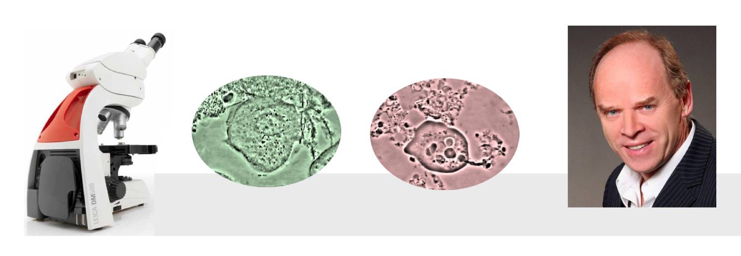
9- To find amoebas, look for the small islands of doughnuts (100x black background) with blackish borders in a field of living neutrophils (exonucleophagy) or an arrangement of moving brush actinomyces (nesting)
10- Note the most relevant or pathogenic results as your main diagnosis
The set-up of the microscope
An optimal setup of the microscope allows the contents of the plate to be adequately visualized.1- LEICA DM microscope 750 eyepieces with 10x widened fields, 10x and 100x Phase lenses, integrated camera
2- Condenser raised to its highest point for maximum contrast
3- Ph3 tab only on the condenser for all operations. This will automatically give you a black background at 100x and a white background at 1000x which is ideal for parasitology research.
4- Keep the condenser surface clean by regularly applying a compress to its surface.
Would you like to be able to apply the full protocol to your patiens?
The seminar "Periodontal Health" will enable you to better understand the theoretical and practical bases of the protocol. At the end of the seminar, you will be able to integrate this approach into your office.The seminar is based on 4 modules, representing the thematic entities that make up the processing:
1. Diagnosis and controls
2. Drug therapy
3. Mechanical therapy
4. Therapeutic education
These modules are interdependent, but a certain autonomy in the respective advancement of each of them is possible, depending on the needs argued by the caregiver and explicitly recorded in the patient's file.
For more information, contact the Institute directly: :
info@parodontite.com